Terms relating to macroscopic morphology, i.e. the shape of veins, are the least consistent, or well defined. Broadly speaking, we can divide veins in two categories:
a) veins that are directly related to some hard object as are pressure fringes (Fig. 2)
b) veins with shapes that are not primarily defined by a relatively
hard object, but by fractures or other factors.
|
|
Figure 2. Pressure fringe of fibrous quartz around a concretion of iron ore in a BIF-chert from the Hamersley ore province, Pilbara, West Australia. Width of view 2.3 mm, crossed polars. |
2.1.1. Pressure fringes
Pressure fringes are veins that form on the two low pressure sides of
hard objects, usually ore minerals, but also other objects, such as crinoid
stems (Durney & Ramsay 1973, Ramsay
& Huber 1983, Selkman 1983, Beutner & Diegel 1985, Etchecopar &
Malavieille 1987, Aerden 1996). They are termed pressure fringes
if they have sharp edges and usually also if their internal structure (see
below) is fibrous (Fig. 2). If not, they are termed pressure shadows (Fig.
3), which usually have diffuse boundaries and not a fibrous internal structure.
One should however note, that recrystallisation can destroy the high grain
boundary energy fibrous internal structure of a pressure fringe, making
it appear like a pressure shadow.
2.1.2. General veins
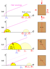
Most veins have the shape of lenses, tabular bodies or blobs. A variety of names (especially in mining; e.g. Barton 1991, Dong et al. 1995) exist for different shapes and positions within the rock. It is impossible to go into details here of every name or term that has been used in the literature, instead, I delineate three broad categories: tension veins, shear veins and breccia veins.
Many veins have their shape and orientation determined by structures
such as fractures, faults or bedding (Fig. 4). The formation of fractures
is favoured by high fluid pressures (Pf), as the differential
stress needed to create fractures is reduced by high fluid pressures (Fig.
5). At sufficiently high differential stress ( Ds
= s 1 - s
3) and Pf, shear fractures form
at angles less than 45° to the maximum stress (Fig. 5.c). Although
some dilation must occur on slip along such fractures, the main mode of
displacement is parallel to the fracture plane and such fractures provide
limited space for vein formation. If the fluid pressure is very high (Pf
> s 3 + T, T = tensile
strength), extensional fractures can form where the main mode of displacement
is normal to the fracture plane (Fig. 5.b) (Secor 1965). Such fractures
provide more space for vein minerals to grow into and indeed, many veins
appear to have grown in such extensional fractures (tension gashes). As
tensional fractures provide the best opportunity for vein formation, it
is not surprising that "tension veins", also called "tension gashes", "tension
fissures" or "gash-veins" are common (Ramsay
& Huber 1983, Rickard & Rixon 1983). These veins are usually
lenticular is shape. Their size can range form mm-size to kilometres (Hippertt
& Massucatto 1998) (Fig. 6). As can be inferred from Fig. 5.b,
the formation of tensional fractures not only requires a high fluid pressure,
but also limits the maximum possible differential stress (Etheridge
1983). Tensile strengths of rocks are generally in the order of
10 MPa, with values reaching several tens of Mpa at the most (Lockner
1995). This limits the differential stress during tensile fracturing
to usually about 20-40 MPa.
Tension veins are often found in en échelon arrays (Fig 7.a).
In such arrays they often have a sigmoidal (S or Z) shape (Durney
& Ramsay 1973, Hanmer 1982, Rickard & Rixon 1983, Selkman 1983,
Nicholson 1991, Olson & Pollard 1991, Passchier & Trouw 1996, Becker
& Gross 1999, Smith 1999). The classical interpretation of such
arrays is simple shearing parallel to the vein array in the direction opposite
the way the vein tips point. The veins originally formed parallel to the
maximum shortening direction (135°) and subsequently rotate. Vein propagation
remains in the 135° direction, resulting in the development of the
sigmoidal shape (Fig. 7.b). New veins may form cutting existing ones and
these veins also initially form in the 135° direction. Continuing deformation
at the en échelon array and formation of new veins in the deformed
zone may eventually lead to the formation of one through-going vein (Fig.
8) (e.g. Wilkinson & Johnston 1996).
|
|
Figure 8. Set of sigmoidal en échelon veins that have amalgamated into a single dextral shear vein. Heavitree Quartzite, Ormiston Gorge, Central Australia. Photograph courtesy Alice Post. |
Whereas tension veins tend to have at least their initial displacement
direction normal to the fracture surface, fault related veins show evidence
for a dominant fault-parallel displacement. Even then, some dilation is
needed to provide space for vein growth. Slickenfibres (Passchier &
Trouw 1996) occur on shear fractures (Fig. 9), but most vein growth is
usually found on more dilatant pull-aparts (Peacock
& Sanderson 1995, Brown & Bruhn 1996). Shear fractures can
form, in intact rocks, at lower Pf than for tensional
fractures, but a higher differential stress is needed (Fig. 5.c). However,
existing planes of weakness (faults, bedding contacts) can reduce the tensional
strength and hence the necessary differential stress and fluid pressure
needed to induce fracturing (Cox &
Paterson 1989, Sibson & Scott 1998) (Fig. 5.d). Veins thus tend
to form along fault planes or parallel to bedding and there particularly
in structures such as folds (e.g. "saddle reefs") and releasing bends (Raybould
1975, Cox et al. 1986, Henderson et al. 1990, Cosgrove 1993,
Jessell et al. 1994, Glen 1995, Windth 1995, Fowler 1996, Fowler
& Winsor 1997, MacKinnon et al. 1997). Another case where
mechanical heterogeneities play an important role in the shape of veins
is that of veins in boudinage necks (Lohest
1909, Cloos 1947, Platt & Vissers 1980, Mullenax & Gray 1984, Malavieille
& Lacassin 1988, Smith, 1998).
|
|
Figure 9. Photograph looking down on slickenfibres in Heavitree Quartzite (Ormiston Gorge, Central Australia). diameter 1 A$ coin approx. 2 cm. Photograph courtesy Alice Post. |
Breccia or net veins form a matrix between clasts in a breccia (Fig.
10). These typically occur in hydrothermal (ore) deposits. True breccia
veins formed in one event of extensive fracturing, without significant
preferred orientation. However, abundant veining of other types and/or
the activity of multiple veining events with different cross-cutting orientations
may produce breccia-like veins (Valenta
et al. 1994).
|
|
Figure 10. Hydrothermal breccia of altered (haematised and silicified) wall rock pieces in a matrix of white quartz. Hammer on right edge of photo for scale. Mt. Gee, Arkaroola, South Australia. |

