2.3.1. Syntaxial veins
In syntaxial veins (Durney & Ramsay 1973)
(Fig. 16.a), growth occurs on a single plane: the median plane. On this
plane, usually a thin fracture, material is added by overgrowth on vein
crystals on both sides of the growth plane. Hence, the latest precipitated
vein material is located at the median plane, while the first and oldest
precipitate is found at the outside of the vein: at the vein - wall rock
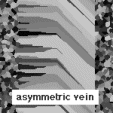
contact. Syntaxial veins can be symmetric, but often the growth plane is
closer to one side of the vein, which produces a marked asymmetry in the
vein (Fig. 16.b) (Fisher & Byrne 1990, Fisher & Brantley 1992).
Syntaxial growth usually occurs when the vein forming mineral is a major
constituent of the host rock. Vein crystals then grow epitaxially off grains
in the wall rock.
2.3.2. Antitaxial veins
When the vein forming mineral is not a major constituent of the host
rock, antitaxial veins (Durney & Ramsay 1973)
commonly form (Fig. 12 & 17). In antitaxial veins there are
two growth surfaces: one on each outer surface of the vein, between vein
and wall rock. New vein material is added simultaneously at both these
surfaces (Fig. 18). Hence, the youngest material is located at the outside
of the vein, while the first precipitate is situated in the middle, on
the median plane. The median plane in an antitaxial vein is clearly of
a different nature than the median plane of a syntaxial vein. In antitaxial
veins, which are usually (always?) fibrous, the median plane is marked
by a string of wall rock inclusions (Fig. 12 & 17) or by a thin zone
of differently textured vein material. Figure 19 shows a case where a zone
of elongate blocky textured calcite occurs in the middle of a fibrous antitaxial
calcite vein.
2.3.3. Composite veins
If a vein is composed of two minerals (e.g. quartz and calcite), it can occur that one mineral shows syntaxial growth, while the other grows simultaneously with an antitaxial growth morphology. The syntaxial mineral started growing from the wall rock inwards, while the second mineral grew from the inside outwards. In this case there are two growth surfaces. Such growth morphologies are called "composite" by Durney and Ramsay (1973). It is suggested here to also use the same term for pressure fringes, and not "complex fringes" as used by Passchier and Trouw (1996). The term composite should be reserved for veins where both morphologies and minerals occupy similar proportions of the vein. Many antitaxial calcite veins, for instance, have a thin rim of syntaxially grown quartz crystals (Fig. 12 & 17) (Williams & Urai 1987, Urai et al. 1991), but these rims are too thin compared to the vein to warrant classification as composite veins.
2.3.4. Syntaxial & antitaxial pressure fringes
The terms syntaxial and antitaxial are also used for pressure fringes
(Durney & Ramsay 1973) (Fig. 20). The
convention for pressure fringes is somewhat confusing as it seems to be
inconsistent with the convention for veins. 'Syntaxial' is a term from
crystallography and denotes overgrowth on a crystal in crystallographic
and mineralogical continuity. Syntaxial veins are called such, as these
veins usually form by crystallographically syntaxial overgrowth of wall
rock grains. 'Antitaxial' (not a crystallographic term) then signifies
the opposite. In an antitaxial veins, the new precipitate is not in crystallographic
or mineralogical continuity with wall rock grains. Instead, growth occurs
by crystallographically syntaxial overgrowth of vein crystals. The terms
syntaxial and antitaxial are therefore defined with respect to a reference
material: the wall rock in case of veins.
It is somewhat confusing at first sight that for pressure fringes, the object was chosen as the reference material and not the wall rock. In a syntaxial pressure fringes, growth occurs on the outside of the object + fringe system and the fringe crystals are syntaxially (or sometimes epitaxially) overgrowing the object. Calcite fringes on a crinoid stems are classical examples of syntaxial pressure fringes (hence the "crinoid-type" of Ramsay & Huber 1983). In an antitaxial pressure fringe, the opposite occurs: the fringe crystals are crystallographically and mineralogically unrelated to the object (and possibly to the wall rock as well). New growth occurs at the contact between object and fringe, with the object often being pyrite ("pyrite-type" of Ramsay & Huber 1983). An excellent overview of the use of pressure fringes for tectonic analysis is given by Passchier & Trouw (1996), who, incidentally, use the term "strain fringe" instead of "pressure fringe". Also note that they use the term "complex fringes" and not "composite fringes" (which would be consistent with Ramsay & Huber 1983) for fringes that exhibit both antitaxial and syntaxial growth.
2.3.5. Ataxial or stretched veins
In the previous cases we saw growth on one or two surfaces. These surfaces
remained the same throughout the growth history. A different class of veins
is formed when the position of the growth surface changes through time
(Fig. 21). This happens when a vein forms from a succession of fractures
that fill with vein material. These fractures can cut the host rock and
vein at different locations and multiple fractures can be present at any
given moment. As such veins are neither syntaxial, nor antitaxial, the
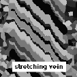
term ataxial is used (Passchier & Trouw 1996). The term 'stretching
veins' (Durney & Ramsay 1973) is also
appropriate, as the resulting grain morphology is that of stretched crystals.
One can recognise two end-member cases of stretching veins: one where all
fractures occur within the growing vein (Fig. 22.a) and one where fractures
occur randomly and it becomes difficult to distinguish between a vein and
the wall rock, as in fact there are multiple veins (Fig. 22.b).
2.3.6. A-syn-anti-bi-uni-taxial?
The terms "syntaxial" and "antitaxial" (Durney & Ramsay 1973) gained
common use in geology thanks to popular text books such as Ramsay &
Huber (1983) and Passchier & Trouw (1996). Passchier & Trouw (1996)
added the term "ataxial" veins for what Ramsay & Huber called "stretching"
veins. They also extended the use of syntaxial and antitaxial to pressure
fringes or strain fringes (see Ch. 2.3.4). More recently, Urai (pers.
comm.) and Hilgers et al. (in press) proposed additional
-taxial terms: "unitaxial" and "bitaxial" (Fig. 23). All these terms may
be confusing if they are not fully and accurately defined and consistently
used by different authors.
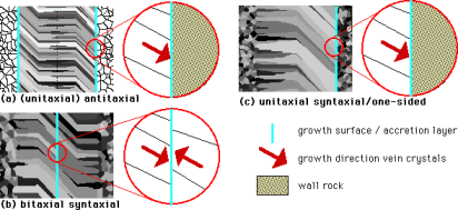
Antitaxial was defined by Durney & Ramsay (1973), Ramsay & Huber (1983) and Passchier & Trouw (1996) for veins in which the crystals inside the vein do grow towards the wall rock, from a median line. In their figures (fig. 13.9 & 13.24 in Ramsay & Huber (1983) and fig. 6.6 in Passchier & Trouw (1996)), antitaxial veins have two growth planes, as in Fig. 12, 17 & 23a. Syntaxial veins grow from and in continuity with the wall rock inwards. Syntaxial veins have only one growth plane (Fig. 16.a and 23.b). At the growth surface of an antitaxial vein, growth is in one direction only and hence they can be termed unitaxial, while growth in syntaxial and stretching veins is from both sides of the growth plane and these veins thus show bitaxial growth.
Whereas the terms "syntaxial", "antitaxial" and "stretching/ataxial" veins refer to the whole vein, the terms "bitaxial" and "unitaxial" refer to growth at one single growth plane. This terminology would normally not cause any problems and one may even argue whether the introduction of the terms "bitaxial" and "unitaxial" is necessary as all antitaxial veins would appear to be unitaxial and all syntaxial and ataxial veins are bitaxial. Problems however arise with completely asymmetric or one-sided veins, that have only one growth plane between the wall rock and one side of the vein (Fig. 16.b and 23.c). Such veins are described by Fisher & Byrne (1990) and Fisher & Brantly (1992) and the "antitaxial fibrous" veins of Cox (1987) could also be such, although it is not fully clear from his figures or text. Are such veins antitaxial because growth is towards the wall rock, albeit only on one side of the vein? Or are they syntaxial, because growth is seeded on the wall rock, albeit only on one side? What is clear, is that these veins are unitaxial, as growth is only in one direction at the growth plane. Ambiguity can be avoided if the terms antitaxial, syntaxial and stretching/ataxial are strictly used to describe a whole vein and unitaxial and bitaxial for the growth situation at a single growth plane:
- An antitaxial vein has two persistent growth planes on the outer surface of the vein, where simultaneous growth occurs unitaxially outwards (Fig. 23.a).
- A syntaxial vein has only one persistent growth plane. Normally growth is bitaxial at that plane and growth is inwards from the wall rock (Fig. 23.b). However, unitaxial growth can occur, in which case the growth plane is on one side between the vein and the wall rock and thus only one half of the syntaxial vein develops (Fig. 23.c).
- Stretching or ataxial veins do not have one or two persistent growth planes but alternating planes at different sites. Growth at the "jumping" growth plane is bitaxial.
2.3.7. Vein morphology and structural analysis
It is clear that syntaxial, antitaxial and composite veins can provide detailed information on the progressive sequence of conditions under which these veins formed. These veins are elongate blocky or fibrous and the direction and curvature of the crystals indicate the kinematics of deformation and the orientation of the stress field. Curvature of vein crystals is a result of progressive change of the vein orientation with respect to the stress field around the vein. Such a change in orientation can result from a change in orientation of the stress field (as for instance in multiple deformation events) and/or a change in orientation of the deforming vein. The correct growth morphology must be determined for any strain analysis. The direction of growth in fibrous or elongate blocky veins can usually be determined by an increase in average grain width in that direction. Careful analysis can reveal this (Durney & Ramsay 1973, Ramsay 1980, Winsor 1987, Passchier et al. 1996, Passchier & Urai 1988, Aerden 1996). It is important in such analyses that it is recognised that crystal shapes do not always fully reflect the displacement path of the vein wall rock, or 'opening trajectory' (Williams & Urai 1987, Urai et al. 1991). A full discussion of the use of veins for structural analysis is beyond the scope of this paper, but this point will be discussed in some more detail in Appendix B.


