Field Relationships
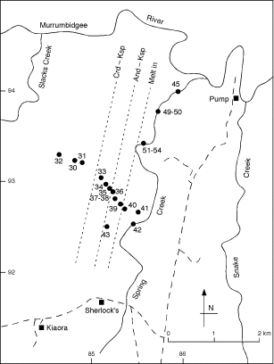 |
Fig. 2 Map showing Spring Creek, Snake Creek and the locations of samples used in this study. |
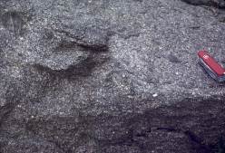 |
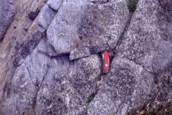
Fig. 3 Typical outcrop appearance of the Cooma Granodiorite, showing euhedral phenocrysts of plagioclase and abundant metasedimentary xenoliths. Pocket knife 9 cm long. |
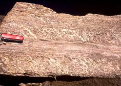
The highest-grade, non-migmatitic rocks are gneisses and granofelses of the andalusite–K-feldspar zone (Fig. 5). The metapsammites have a strong differentiated layering, which has led to their being called "corduroy gneisses" (e.g., Joplin, 1942), whereas the metapelites are coarse-grained and studded with porphyroblasts of cordierite, many of which have been partly to completely replaced by fine-grained, symplectic aggregates of biotite, andalusite and quartz (Vernon, 1978). These dark pseudomorphs are why the metapelites have been called "mottled gneisses" (e.g., Joplin, 1942).
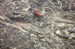
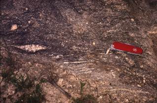
In contrast to the situation at low and medium metamorphic grades, the high-grade metapelites are stronger than the interbedded metapsammites, owing to their abundant coarse-grained, strong minerals, especially cordierite, andalusite and K-feldspar. The result is that the metapsammites commonly are folded and foliated in an intricate manner, whereas the metapelites tend to fracture and undergo boudinage (Figs 6, 7). This fracturing appears to have assisted migration of melt, as discussed later.
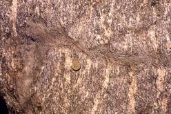 |
Fig. 7 Magnified view of part of Fig. 6, showing the intense deformation of a metapsammite bed. |
The first indications of partial melting with increasing metamorphic grade are rare, local, small patches of leucosome in the metapelites, grading into veinlets (e.g. sample 40). These veinlets have been crenulated by the main foliation, S3, applying the foliation nomenclature of Johnson & Vernon (1995a). Therefore, they pre-date or formed during the development of S3. A few leucosome veinlets are larger and coarser-grained, some being localized on metapelite-metapsammite contacts (Fig. 8). At slightly higher grades (e.g. sample 41), the leucosome is concentrated in small lenses parallel to S2 and S3, as well as along S0, and is folded by F3 folds. In Spring Creek (e.g. sample 42), the main migmatite zone is entered and the above relationships are well developed.
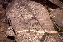 |
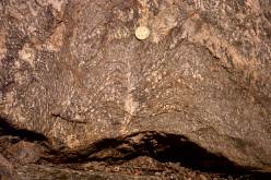
Fig. 8 Leucosome concentrated at the junction of metapelite (with porphyroblasts of cordierite replaced by dark symplectite) and metapsammite beds, Spring Creek. Coin 2.5 cm in diameter. |
Prominent lenses and veins of white leucosome are oblique to S3. These veins (Figs 9) show a strong tendency to be parallel throughout the Spring Creek-Snake Creek area (Fig. 2). We interpret them as having formed during D3 by fracturing along S2, together with incipient boudinage of the strong metapelitic beds, providing low-pressure sites for local segregation of partial melt. The fact that these leucosome veins generally do not follow the more prominent S3 is consistent with fracturing during D3, because if the direction of maximum finite elongation was parallel to S3 during D3, fracturing should have occurred along surfaces at moderate to high angles to S3. Earlier leucosomes, formed before or early during the development of D3 , tend to be folded into parallelism with S3 (Fig. 9).
In some metapsammites, boudinage of thin metapelitic layers — M domains in a differentiated S3 ("corduroy") layering — has occurred, with leucosomes in the interboudin zones (Fig. 10). This suggests that the leucosomes formed late during D3.
The leucosomes also have a strong, though more local tendency to concentrate in the metapelites along the bases of metapsammite beds (Fig. 8), presumably owing to opening of fractures along this zone of inferred competency contrast. Some of these leucosomes have been crenulated by S3, indicating their formation before or early during D3. This is mainly seen in the hinges of parasitic F3 folds, where bedding is at high angles to the direction of maximum finite elongation (S3) during D3(Fig. 9). Even where bedding is not at a high angle to S3 now, presumably it was at the onset of D3, because F3 folds control the macroscale structural geometry in this area (Hopwood, 1976; Johnson et al., 1994).
A prominent feature that persists through most of the migmatites at Cooma is the strong tendency for the confinement of leucosome to metapelitic beds, so that the rocks resemble the "bedded migmatites" of Greenfield et al. (1997), as shown in Fig. 11. Locally, leucosomes penetrate adjacent metapsammitic beds.
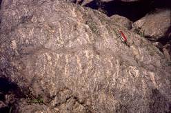
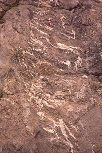
Most of the Cooma leucosomes consist only of quartz and K-feldspar, though porphyroblasts of cordierite are fairly common and pink andalusite occurs locally, e.g., in Spring Creek. Melanosomes are rare. The amount of partial melting increases to a maximum in Snake Creek and further east (Figs 1, 2), the migmatites tending to become coarser-grained and more stromatic (Figs 12, 13, 14, 15), though remaining essentially "bedded migmatites" (Fig. 12). The stromatic character is more prominent further to the east, e.g., in Pilot Creek (Figs 14. 15), which may be related to the stronger deformation that these rocks appear to have undergone. Folded remnants of metapsammite occur in the most extensively melted migmatites (Fig. 15). Many of the metapelites in Snake Creek contain abundant retrograde muscovite, and pegmatitic aggregates are patchy to pervasive, many containing acicular to prismatic tourmaline. These features suggest late access of water.
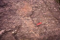 |
Fig. 12 Coarser grained and more completely developed leucosome, but still confined to metapelite beds, Snake Creek (Fig. 1). Pocket knife 9 cm long. |
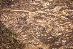 |
Fig. 13 Intensely deformed, folded, stromatic migmatite, Snake Creek. Coin 2.5 cm in diameter. |
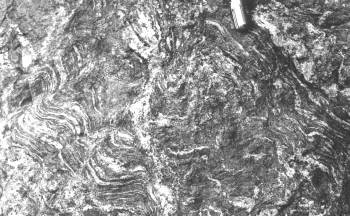 |
Fig. 14 Intensely folded stromatic migmatite, with leucosomes occurring in a fold axial surface (centre), Pilot Creek, between Snake Creek and Cooma Creek (Fig. 1). Pocket knife 9 cm long. |
Locally, patches and small, transgressive to sheet-like intrusions of plagioclase-bearing microgranitic material, with small rectangular crystals of plagioclase, occur in the highest-grade migmatites (e.g. in Snake Creek), typically with scattered metasedimentary xenoliths and migmatite remnants similar to those in the Cooma Granodiorite (Fig. 16). This plagioclase-bearing leucosome has disrupted and boudinaged the metapelite leucosomes (Fig. 17), and shows only the latest foliation (S5), as does the Cooma Granodiorite. It appears to have originated by partial melting of quartzofeldspathic metasediments (psammitic or psammopelitic), as leucosomes of this type commonly occur in these rocks in Snake Creek, and rarely in Spring Creek. Therefore, the plagioclase-bearing leucosome is a potential source for the Cooma Granodiorite magma, although this hypothesis requires testing by mineralogical and chemical investigations, which are in progress.
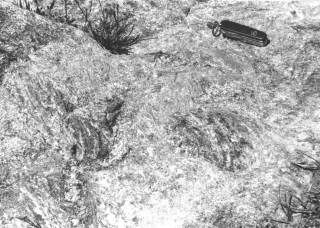 |
Fig. 16 Large patch of intrusive material resembling Cooma Granodiorite, containing migmatite xenoliths, Snake Creek (Fig. 1). Pocket knife 9 cm long. |
