Microstructural Relationships
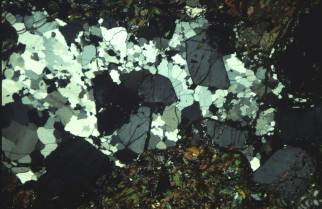
(1) Inclusion trails are absent, in contrast to grains of the same minerals in the mesosome (Figs 19, 20).
(2) Overgrowths free of inclusions trails may occur on minerals (e.g., K-feldspar, cordierite) with inclusion trails, as shown in Fig. 19.
(3) Crystal faces of K-feldspar (and cordierite?) may occur against quartz (e.g., Vernon & Collins, 1988), as shown in Figs 19 and 20.
(4) Simple twinning may occur in K-feldspar, which appears to be diagnostic of crystallization of K-feldspar in a melt, rather than the solid state (Vernon, 1986, Vernon, 1998).
 |
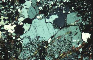
Fig. 20 Deformed leucosome in Snake Creek, showing euhedral K-feldspar (K), which has resisted the deformation and quartz (Q), which has recrystallized. Crossed polars; base of photo 1.5 mm. |
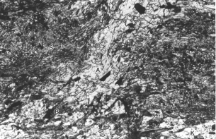
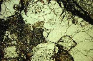
Leucosome first appears in the metapelites (sample 40), in which it forms small, clear patches (a few millimetres across) and short veinlets that contrast strongly with the mesosome, in which the cordierite and K-feldspar are full of inclusions (Fig. 21). The initial leucosome patches are so small that they are not obvious in the field, but larger veinlets appear at slightly higher grade (metapelite sample 41), just west of Spring Creek. The initial leucosome is accompanied by the first appearance of fibrous sillimanite as crenulated and contorted, discontinuous folia (sample 40); this sillimanite is different in appearance and timing from the late sillimanite that has replaced all minerals, especially along grain boundaries, in some of the high-grade rocks at Cooma (Vernon 1979).
In thin section, leucosomes in the Cooma metasediments are observed to consist mainly of quartz and microperthitic K-feldspar, with some cordierite, rarely with minor biotite or andalusite; plagioclase grains were not observed, although rare plagioclase in metapelite leucosome was reported by Ellis & Obata (1992, p. 101). As noticed in the field, the leucosomes observed in thin section do not have melanosomes, suggesting that either (i) the leucosomes have moved away from sites of initial segregation, leaving behind concentrations of mafic aggregates, or (ii) the cordierite produced in the biotite dehydration melting reaction (see later) was localized as porphyroblasts, by growing on existing cordierite grains, rather than growing as a continuous fringe on the leucosome. Thus, the patchy leucosomes are probably in contact with residual mafic minerals, as indicated by cordierite with inclusion trails projecting into inclusion-free cordierite in leucosome.
Some of the cordierite in the leucosomes shows evidence of replacement by symplectic aggregates of biotite, quartz and andalusite, which is very common in the mesosomes and non-migmatitic metapelitic gneisses (Vernon, 1978). However, some is unaltered, as reported for the outcrop studied by Ellis & Obata (1992). Back-reaction of cordierite to biotite as the leucosome melt cooled would be expected, and Ellis & Obata (1992) suggested that the absence of back-reaction is due to extraction of hydrous melt from the leucosome. It probably would be difficult to avoid at least some back-reaction during this process, and a possible alternative explanation is that armouring of the cordierite by the first quartz and K-feldspar to crystallize from the melt (i.e., by heterogeneous nucleation on the cordierite) prevented reaction. The water expelled from the cooling leucosomes could have entered the adjacent rocks and reacted with the cordierite to produce the abundant symplectite (Vernon, 1978; Vernon & Pooley, 1981), as suggested for cordierite in metapelites at Mount Stafford, central Australia, by Vernon et al. (1990).


